Research Interests
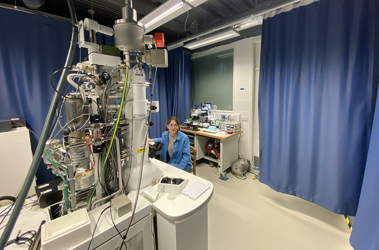
Cells are continuously challenged by a variety of dangerous cytosolic materials, including invading bacteria, misfolded proteins or damaged organelles. All of these need to be promptly detected, triaged and possibly cleared to ensure maintenance of physiological functions and cell survival. If these pathways fail, the consequences are catastrophic: accumulation of protein aggregates is associated with neurodegenerative diseases such as Alzheimer’s and Parkinson’s; impaired lipid turnover contributes to metabolic syndromes including type 2 diabetes and fatty liver disease; and defective organelle recycling can trigger inflammatory disorders and compromise immune responses. In my current work, I use bacterial infection models, combined with structural biology and biochemical methods, to understand how cells defend themselves against cytosol-invading bacteria. At the same time, it is becoming apparent that the same pathways that survey the cytosol for invading pathogens are also repurposed for danger sensing and resolution in many other physiological contexts. Therefore, I am interested in elucidating the universal and cell-type specific molecular principles that enable cells to detect diverse threats and decide their fate - destroy, recycle, or tolerate. Structural biology is a powerful tool to investigate these mechanisms, but putting protein structures into their cellular context remains challenging. Building on my background in physics and on my previous experience in methods development for electron microscopy (see my Publications page for more details), I continue to design new methods and instruments that extend the limits of what we can see inside cells, enabling experiments that are impossible with existing technologies.
Research Highlights
HexAuFoil: improved specimen supports for cryoEM, that eliminate specimen movement and can be manufactured at scale.
Structure of the light-harvesting complex from the bacterium Gemmatimonas phototrophica.
Visualisation of the effects of radiation damage on protein structure during imaging with electrons.
A wafer-scale, customisable process for cryoEM grid manufacturing, inspired by techniques used in the semiconductor industry.
A low-cost 100 kV cryogenic electron microscope, designed specifically for protein structure determination.
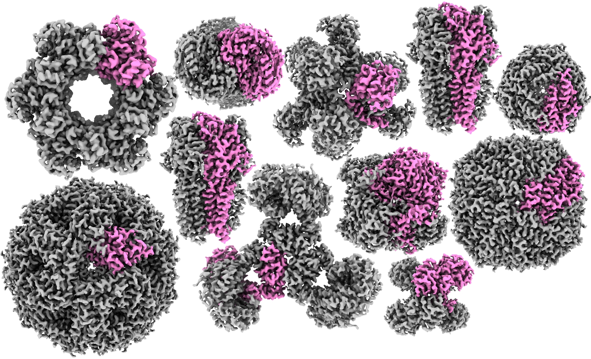
A collection of protein structures, determined using cryoEM at 100 keV.
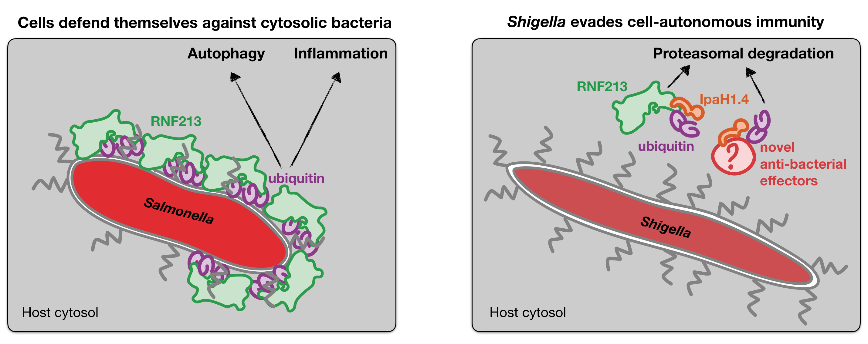
Illustration of the host-pathogen arms race, where the host protein RNF213 defends the cytosol from invading bacteria by ubiquitylating them, whereas certain cytosol-adapted bacteria, such as Shigella flexneri have evolved effector proteins that ubiquitylate and degrade the host anti-bacterial effectors.
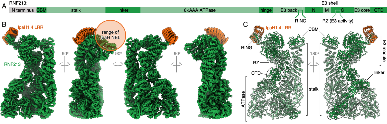
Structure of the host protein RNF213 bound to the bacterial secreted effector IpaH1.4, that can target RNF213 for degradation.
Publications
A complete and up-to-date list of my publications can be found on my Google Scholar page.
- JL Dickerson, K Naydenova, MJ Peet, H Wilson, B Nandy, G McMullan, R Morrison & CJ Russo. Reducing the effects of radiation damage in cryo-EM using liquid helium temperatures. PNAS (2025).
- K Naydenova*, K Boyle*, C Pathe*, P Pothukuchi, A Crespillo-Casado, F Scharte, P-M Hammoudi, EG Otten & F Randow. Shigella flexneri evades LPS ubiquitylation through IpaH1.4-mediated degradation of RNF213. NSMB (2025).
- Y Hooda*, A Sente*, RM Judy*, L Smalinskaitė, S-Y Peak-Chew, K Naydenova, T Malinauskas, SW Hardwick, DY Chirgadze, AR Aricescu & RS Hegde. Mechanism of NACHO-mediated assembly of pentameric ligand-gated ion channels. bioRxiv (2024, preprint).
- A Crespillo-Casado*, P Pothukuchi*, K Naydenova, MCJ Yip, JM Young, J Boulanger, V Dharamdasani, C Harper, P-M Hammoudi, EG Otten, K Boyle, M Gogoi, HS Malik & F Randow. Recognition of phylogenetically diverse pathogens through enzymatically amplified recruitment of RNF213. EMBO Reports (2024).
- G McMullan, K Naydenova, D Mihaylov, K Yamashita, MJ Peet, H Wilson, JL Dickerson, S Chen, G Cannone, Y Lee, KA Hutchings, O Gittins, MA Sobhy, T Wells, MM El-Gomati, J Dalby, M Meffert, C Schulze-Briese, R Henderson & CJ Russo. Structure determination by cryoEM at 100 keV. PNAS (2023).
- JL Dickerson, E Leahy, MJ Peet, K Naydenova & CJ Russo. Accurate magnification determination for cryoEM using gold. Ultramicroscopy (2023).
- CJ Russo, JL Dickerson & K Naydenova. Cryomicroscopy in situ: what is the smallest molecule that can be directly identified without labels in a cell? Faraday Discussions (2022).
- K Naydenova, A Kamegawa, MJ Peet, R Henderson, Y Fujiyoshi & CJ Russo. On the reduction in the effects of radiation damage to two-dimensional crystals of organic and biological molecules at liquid-helium temperature. Ultramicroscopy (2022).
- K Naydenova & CJ Russo. Integrated wafer-scale manufacturing of electron cryomicroscopy specimen supports. Ultramicroscopy (2022).
- A Sente, R Desai, K Naydenova, T Malinauskas, Y Jounaidi, J Miehling, X Zhou, S Masiulis, SW Hardwick, DY Chirgadze, KW Miller & AR Aricescu. Differential assembly diversifies GABAA receptor structures and signalling. Nature (2022).
- P Qian, AT Gardiner, I Šímová, K Naydenova, TI Croll, PJ Jackson, Nupur, M Kloz, P Čubáková, M Kuzma, Y Zeng, P Castro-Hartmann, B van Knippenberg, KN Goldie, D Kaftan, P Hrouzek, J Hájek, J Agirre, CA Siebert, D Bína, K Sader, H Stahlberg, R Sobotka, CJ Russo, T Polívka, CN Hunter & M Koblížek. 2.4 Å structure of the double ring Gemmatimonas phototrophica photosystem. Science Advances (2022).
- K Naydenova*, KW Muir*, L-F Wu*, Z Zhang, F Coscia, MJ Peet, P Castro-Hartmann, P Qian, K Sader, K Dent, D Kimanius, JD Sutherland, J Löwe, D Barford & CJ Russo. Structure of the SARS-CoV-2 RNA-dependent RNA polymerase in the presence of favipiravir-RTP. PNAS (2021).
- AT Gardiner*, K Naydenova*, P Castro-Hartmann, TC Nguyen-Phan, CJ Russo, K Sader, CN Hunter, RJ Cogdell & P Qian. The 2.4 Å cryo-EM structure of a heptameric light-harvesting 2 complex reveals two carotenoid energy transfer pathways. Science Advances (2021).
- K Naydenova, P Jia & CJ Russo. Cryo-EM with sub–1 Å specimen movement. Science (2020).
- K Naydenova, G McMullan, MJ Peet, Y Lee, PC Edwards, S Chen, E Leahy, S Scotcher, R Henderson & CJ Russo. CryoEM at 100 keV: a demonstration and prospects. IUCrJ (2019).
- K Naydenova*, MJ Peet* & CJ Russo. Multifunctional graphene supports for electron cryomicroscopy. PNAS (2019).
- K Naydenova & CJ Russo. Measuring the effects of particle orientation to improve the efficiency of electron cryomicroscopy. Nature Communications (2017).
Brief Bio
During my high-school years, I was an avid competitor in the International Physics Olympiads, which secured me a place to read the Natural Sciences Tripos (BA and MSci) at the University of Cambridge, specialising in physics. During this time, I first joined Chris Russo's group in the Structural Studies Division of the MRC Laboratory of Molecular Biology (MRC LMB) in Cambridge as a summer student in 2016. We were having a great time, so I stayed on for another summer internship, as well as for my master's project, my PhD and a brief postdoc. We elucidated the physical principles that limit how well one can image biological molecules and we built technologies to improve the capabilities of cryogenic electron microscopy (cryoEM) for structural biologists: for example, during my PhD I developed the HexAuFoil cryoEM grids. For this work, I was awarded the Max Perutz Prize for outstanding PhD research in 2020. By the end of my PhD, in 2023, the cryoEM field was growing exponentially and hundreds of protein structures were being solved per week - and I realised it was time for a new adventure. So for my postdoc, I moved into cell biology, and joined the group of Felix Randow, also at the MRC LMB. Our group studies cell-autonomous immunity, which is the ability of non-specialised, non-immune cells to defend themselves against pathogens. We also investigate how professional cytosol-dwelling pathogens evade these host defences. Alongside my research, I also enjoy teaching at the annual Cold Spring Harbor cryoEM course in the USA.
Academic History
2023 – 2025 Career Development Fellow. Investigation of anti-bacterial innate immune signalling pathways. Felix Randow group in the PNAC Division of MRC LMB, Cambridge, UK.
2022 – 2023 Postdoctoral Fellow. Development of new methods for in situ cryogenic electron microscopy. Chris Russo group in the Structural Studies Division of MRC LMB, Cambridge, UK.
2018 – 2022 PhD in Biological Sciences. Thesis: The physical origins of specimen movement in electron cryomicroscopy and how to eliminate it. Chris Russo group in the Structural Studies Division of MRC LMB, Cambridge, UK.
2017 – 2018 MSci in Natural Sciences (Physics) from the University of Cambridge. Thesis: Spectroscopy of genetically encodable fluorescent proteins at cryogenic temperatures. Chris Russo group in the Structural Studies Division of MRC LMB, Cambridge, UK.
2016 – 2017 Research Associate (Summer Intern). Development of methods for determining, controlling, and analysing the effects of the surface energy of cryoEM specimen supports. Chris Russo group in the Structural Studies Division of MRC LMB, Cambridge, UK.
2014 – 2017 BA Hons in Natural Sciences (Physics) from the University of Cambridge, UK.
Resources
Talks and Lectures
CryoEM specimen preparation. This lecture is part of the 2023 MRC LMB CryoEM course, which is intended to be watched as a continuation of the previous years' courses.
New specimen support technologies for cryoET. I gave this talk at the Chan Zuckerberg Imaging Institute CryoET Hardware Workshop in Redwood City, CA, USA in May 2023. Here, I summarise everything we have learned about the physics of cryoEM specimens in the context of isolated proteins, and how this knowledge might be extended to cellular specimens and electron tomography.
Molecular structure extrapolation to zero dose with cryoEM. I gave this talk at the Institute for Pure and Applied Mathematics in UCLA, USA in November 2022. I describe how we developed a method to acquire movement-free cryoEM micrographs, and how this, in turn, has allowed us to develop a computational method for calculating the cryoEM structures of undamaged biological molecules.
Press
- Cambridge Independent: New cryo-EM frontier opens at -260C thanks to research at MRC Laboratory of Molecular Biology in Cambridge
- MRC LMB Insight on Research: Cytosol-adapted bacterium usurps host defence mechanism to evade LPS ubiquitylation
- Microscopy Today: HexAuFoil cryo-EM specimen support grids named as a winner of the 15th Microscopy Today Innovation Awards
- Cambridge Independent: New era of lower-cost, lower-energy cryo-EM ushered in by MRC Laboratory of Molecular Biology in Cambridge
- MRC LMB Insight on Research: Low energy cryo-EM to widen accessibility for fast and accurate structure determination
- Science: Cheaper microscope could bring protein mapping technique to the masses
- Nature: A low-cost electron microscope maps proteins at speed
- Phys.org: Researchers design smaller, more cost-efficient electron cryo-microscope
- Diamond Light Source News: Scientists show how an enigmatic bacterium from the Gobi desert harvests solar energy
- MRC LMB News: LMB students recognised by Cambridge Society for the Application of Research
- MRC LMB Insight on Research: Scaling-up grid manufacture to solve bottleneck in cryo-EM
- Cambridge Independent: Global bottleneck holding up discovery using cryo-EM is solved by MRC Laboratory of Molecular Biology in Cambridge
- MRC LMB Insight on Research: Targeting RNA replication in SARS-CoV-2
- MRC LMB Insight on Research: Freezing molecules completely still for cryo-EM
- Science: Better, faster, and even cheap
- Wiley Analytical Science: New grid minimizes the movement problem in cryo-EM
- Phys.org: Support film makes cryo-electron microscopy sharper
- MRC LMB News: Student and postdoc prizes awarded for outstanding work
- Medical Xpress: Slashing energy could make cryoEM microscopes affordable for many labs worldwide
- Cambridge Independent: LMB unveils vision for next generation of Nobel Prize-winning cryo-EM technology
- MRC LMB Insight on Research: Fast, simple, accessible and affordable: The future of cryo-EM
- MRC LMB Insight on Research: Functionalized graphene sheets on gold grids aid structure determination by electron cryo-microscopy
Contact Details
Email me at katerinanaydenova [at] gmail [dot] com